When an X-Ray Alone Isn’t Enough: Next Steps in Imaging
We’ve all been there: a clumsy fall, a persistent ache, a nagging concern that something just isn’t right. The first stop for many of us is an X-ray. It’s a quick, painless way to get a look inside your body and see if you’ve broken a bone. And often, that’s all it takes. The doctor can confirm a fracture, a dislocation, or show that everything is perfectly fine.
But what happens when the X-ray results are inconclusive? Or when your symptoms point to a soft tissue injury that an X-ray can’t even see? This is where the world of medical imaging opens up, revealing a host of powerful tools that go far beyond a simple X-ray.
The Limitations of an X-Ray
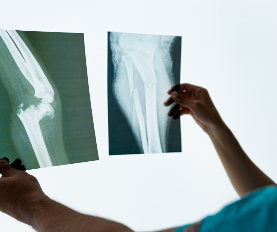
An X-ray is a fantastic diagnostic tool, but it has its limits. Think of it as a snapshot of your bones. It uses electromagnetic radiation to create a two-dimensional image, with dense materials like bone appearing white and softer tissues like muscle and fat appearing in shades of gray.
While this is perfect for detecting fractures and certain bone abnormalities, it’s not designed to visualize:
- Soft Tissues: This includes muscles, tendons, ligaments, and cartilage. A torn ACL, a rotator cuff injury, or a herniated disc won’t show up on an X-ray.
- Detailed Organ Structures: While an X-ray can show the general shape of organs, it can’t provide the detailed, cross-sectional view needed to diagnose conditions like tumors, cysts, or internal bleeding.
- Early-Stage Conditions: Some bone-related issues, like stress fractures or early signs of arthritis, might not be immediately visible on an X-ray.
So, if your doctor says the X-ray is clear but your pain persists, what’s next?
Beyond the X-Ray: Your Guide to Advanced Imaging
Once an X-ray has been ruled out as the final word, your doctor will likely recommend one of these more advanced imaging techniques. Each one offers a different perspective and is suited for specific diagnostic needs.
1. MRI (Magnetic Resonance Imaging)
- How it works: An MRI uses powerful magnetic fields and radio waves to create detailed, three-dimensional images of the body’s soft tissues. It doesn’t use radiation, making it a safe option for repeated use.
- What it’s good for: This is the go-to for soft tissue injuries. Think torn ligaments in a knee (ACL, MCL), a herniated disc in the spine, or a rotator cuff tear in the shoulder. It’s also invaluable for diagnosing neurological conditions, such as multiple sclerosis (MS), and for examining organs like the heart, liver, and brain.
- The experience: You’ll lie on a table that slides into a large, tube-like machine. It can be a little noisy, and some people find the enclosed space uncomfortable, but you can often request an open MRI machine if you’re claustrophobic.
2. CT Scan (Computed Tomography)
- How it works: A CT scan uses a series of X-ray images taken from different angles to create cross-sectional, detailed images of bones, blood vessels, and soft tissues. It’s like taking a loaf of bread and looking at each individual slice.
- What it’s good for: CT scans are excellent for visualizing complex bone fractures, especially in areas like the pelvis or spine. They are also crucial in emergency situations to quickly assess internal injuries, bleeding, or strokes. They can also be used to detect tumors and other abnormalities in organs.
- The experience: Similar to an MRI, you’ll lie on a table that moves through a doughnut-shaped machine. The scan is typically much faster than an MRI.
3. Ultrasound
- How it works: Ultrasound uses high-frequency sound waves to create real-time images of the inside of the body. There is no radiation involved.
- What it’s good for: Most people associate ultrasound with pregnancy, but its uses are far broader. It’s excellent for examining a wide range of soft tissues, including tendons, muscles, and ligaments, often used to diagnose a muscle tear or a tendon rupture. It’s also the standard for viewing abdominal organs, the thyroid gland, and blood flow.
- The experience: A technician will apply a special gel to your skin and then press a small transducer device against the area being examined. You can often see the images on a screen in real time.
Getting the Right Diagnosis is Key
When an X-ray doesn’t provide the answers, it’s not a dead end. It’s simply the first step on the path to a more precise diagnosis. Your doctor will carefully consider your symptoms, medical history, and the initial X-ray results to determine which advanced imaging test is the most appropriate next step.
By understanding the unique capabilities of each of these imaging techniques—MRI, CT scan, and ultrasound—you can feel more informed and prepared for your next appointment. Getting a clear and accurate diagnosis is the most important step toward getting the right treatment and starting your journey to recovery.
No comment