
Accidents happen when you least expect them—a sudden trip on the pavement, an awkward landing on the sports field, or a minor slip at home. While minor bumps and bruises usually heal with rest and ice, deeper structural injuries require a clearer look under the surface.
Medical imaging, specifically diagnostic digital radiography (X-rays), allows healthcare providers to visualize bones, joints, and surrounding internal structures without invasive procedures. Knowing when an injury warrants medical imaging can save you from delayed healing, chronic pain, or long-term joint complications.
Here are 5 common injuries where getting an X-Ray is crucial for an accurate diagnosis and proper recovery plan.
1. Bone Fractures (Broken Bones)
Bone fractures are the most frequent reason doctors order an X-ray. Whether caused by sudden high-impact trauma, a fall, or repetitive stress over time, fractures aren’t always obvious visible breaks through the skin.
- Full Fractures: High-energy impacts from sports or vehicle collisions can cause clean breaks or shattered bone fragments. An X-ray reveals the exact angle and displacement of the break so physicians can properly align (reduce) the bone.
- Stress Fractures: Common among runners and athletes, stress fractures are micro-cracks caused by repetitive force. While subtle, X-rays help confirm bone remodeling or structural cracks when localized pain persists.
- Hairline Fractures: Tiny cracks in bones—frequently in the feet, shins, or wrists—can feel like severe bruising or deep aching. Without an X-ray, hairline fractures can easily be misdiagnosed as simple muscle strain, leading to worsening injury.
2. Joint Dislocations
A dislocation occurs when extreme force pops the bones forming a joint out of their normal anatomical alignment. The most frequently dislocated joints include shoulders, fingers, knees, and elbows.
An X-ray serves two vital purposes during a dislocation:
- Pre-Reduction Assessment: Confirms the direction and extent of the dislocation while checking if any small bone chips broke off during the displacement (avulsion fracture).
- Post-Reduction Verification: Once a doctor manually maneuvers the joint back into place, a follow-up X-ray verifies that the bones are seated correctly within the joint capsule.
3. Severe Sprains and Ligament Tears
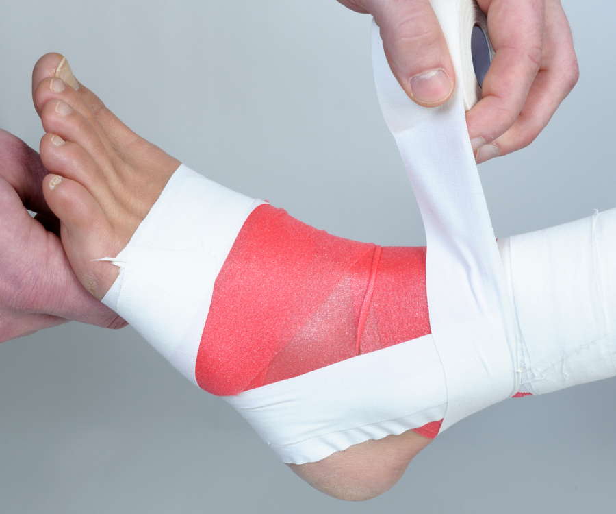
When you roll an ankle or twist a knee, the primary damage is often to ligaments—soft tissue that connects bone to bone. While standard X-rays do not directly image soft tissue like ligaments or tendons (which are better evaluated via MRI or ultrasound), an X-ray is almost always the first step in acute joint trauma.
Physicians use clinical guidelines like the Ottawa Ankle Rules to determine if an X-ray is necessary. The primary goal is to rule out hidden avulsion fractures—where a tearing ligament pulls off a tiny piece of attached bone—and ensure the joint alignment remains stable under stress.
4. Chest Wall Trauma and Rib Injuries
A hard blow to the chest from a fall, steering wheel impact, or contact sport can cause severe localized chest pain that hurts worse with deep breathing, coughing, or laughing.
Chest and rib X-rays are critical not just to check for cracked or fractured ribs, but to evaluate potential underlying complications:
- Pneumothorax: A fractured rib tip can puncture a lung, causing air to leak into the chest cavity and collapse the lung.
- Hemothorax: Blood pooling in the pleural cavity around the lungs following blunt trauma.
- Internal Organ Bruising: Visualizing the rib cage gives emergency doctors vital context regarding the force delivered to the torso.
5. Foreign Body Ingestion or Deep Penetration Trauma
Accidental ingestion or puncture wounds involving dense foreign materials often require immediate X-ray imaging.
- Ingested Objects: Children (and occasionally adults) accidentally swallow coins, small metal battery cells, or hardware. An X-ray identifies the exact location of radiopaque objects in the esophagus, stomach, or intestinal tract to determine if endoscopic removal is necessary.
- Deep Puncture Wounds: Glass shards, metallic splinters, or gravel buried deep inside a puncture wound can cause severe infection or tissue damage if left behind. An X-ray highlights radiopaque debris so surgeons can locate and safely extract every piece.
Key Symptoms: When Should You Seek an Immediate X-Ray?
Not every injury requires an emergency scan, but you should visit an urgent care clinic or emergency room for diagnostic imaging if you notice any of these warning signs:
| Symptom | What It Indicates |
| Inability to bear weight | Inability to take 4 steps on an injured foot, ankle, or leg often signals a fracture. |
| Visible bone deformity | Crooked fingers, limbs resting at unnatural angles, or abnormal joint humps. |
| Severe pinpoint tenderness | Intense, sharp pain directly over a bone rather than surrounding muscle. |
| Numbness or tingling | Tingling in toes or fingers suggests bone fragments or joint pressure on nerves. |
| Immediate severe swelling | Rapid bruising and localized tight swelling within minutes of the injury. |
What to Expect During Your X-Ray Procedure
Getting an X-ray is quick, painless, and non-invasive. The radiological technologist will position the injured body part against an imaging plate or table. You will be asked to remain completely still for a few seconds while the machine emits a brief beam of low-dose radiation through the tissue. Dense materials like bone absorb the radiation, appearing white on the final digital image, while soft tissues appear in shades of gray.
No comment