
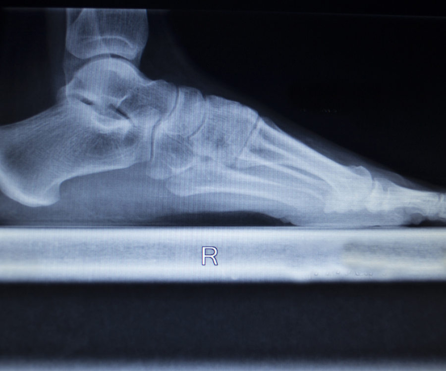
A weight-bearing X-ray (also called a standing X-ray) is a diagnostic imaging test taken while you support your full body weight on the area being evaluated—most commonly your feet, ankles, knees, hips, or spine.
Unlike standard non-weight-bearing radiographs taken while lying on an examination table, weight-bearing views reveal how gravity, joint space narrowing, and mechanical stress affect skeletal alignment during everyday movement.
Why Doctors Order Weight-Bearing Radiographs
When you sit or lie down, your muscles and tendons relax, and the pressure inside your joint capsules drops. This often causes bones to shift back into a “neutral” position, masking underlying structural deficits.
- Assessing Cartilage Loss & Joint Space: In conditions like knee osteoarthritis, cartilage thinning is only clearly visible when the femur compresses down onto the tibia under body weight.
- Evaluating Foot & Ankle Alignment: Issues like flat feet (pes planus), bunions (hallux valgus), and Lisfranc ligament sprains become pronounced under load.
- Spinal Deformity Mapping: Spinal conditions such as scoliosis and spondylolisthesis shift noticeably between supine (lying flat) and upright postures.
- Surgical & Orthotic Planning: Orthopedic surgeons rely on weight-bearing views to accurately plan total knee arthroplasties (TKR), ligament reconstructions, and custom orthotic devices.
Weight-Bearing vs. Standard Non-Weight-Bearing X-Rays
| Feature | Weight-Bearing X-Ray | Non-Weight-Bearing X-Ray |
| Patient Position | Standing upright, bearing full or partial weight | Lying supine, prone, or seated |
| Primary Focus | Functional alignment, joint space narrowing, stability under load | Acute trauma, bone fractures, gross bone morphology |
| Key Clinical Uses | Osteoarthritis, foot arch collapse, subtle ligament instability | Non-ambulatory patients, severe trauma, bed-bound diagnostics |
| Mechanical Stress | Realistic functional load | Zero joint compression |
What to Expect During the Procedure
The procedure is quick, non-invasive, and uses low-dose radiation:
- Positioning: You will stand on an elevated platform or directly on the floor adjacent to the X-ray detector plate.
- Weight Distribution: The radiographer will instruct you to distribute your weight evenly across both legs, or temporarily shift your full weight onto the injured limb for specific views.
- Safety & Stability: Support handles are provided if standing causes pain or instability.
- Image Capture: You hold still for a few seconds per view while the technician captures the scans from anterior (front), lateral (side), or specialized angled projections.
If severe pain prevents you from standing unassisted, notify the technologist before the scan so they can provide supportive aids or discuss modified partial weight-bearing views with your referring physician.
No comment