
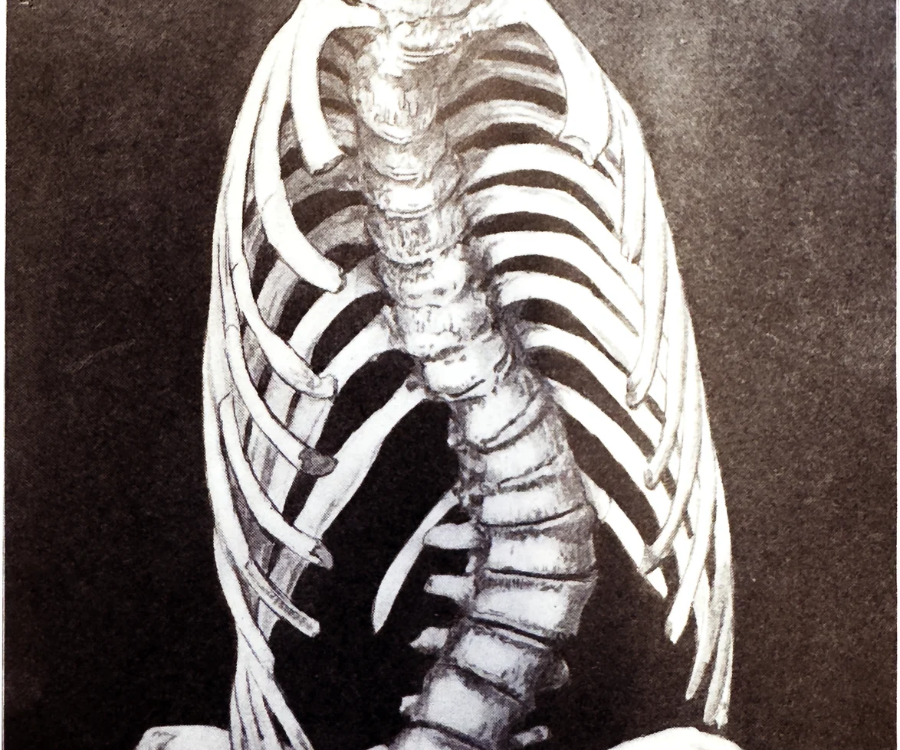
Scoliosis in Teenagers: How Can X-Rays Help?
Noticing uneven shoulders, a protruding shoulder blade, or a tilted waistline in a teenager often brings an immediate wave of questions for parents. While a physical screening—such as the classic forward-bend test—can highlight physical asymmetries, doctors cannot see what is happening beneath the skin without diagnostic imaging.
When evaluating scoliosis in teenagers, spinal X-rays serve as the clinical gold standard. They transform a suspected curve into precise, measurable data, allowing orthopedic specialists to diagnose the condition accurately, track progression during growth spurts, and build an effective treatment plan.
Why X-Rays Are the Gold Standard for Adolescent Scoliosis
Teenage scoliosis—most commonly adolescent idiopathic scoliosis (AIS)—typically emerges between ages 10 and 18, right as the skeleton undergoes rapid growth. Because bones are still developing, a mild curve can remain stable or progress rapidly within a few months.
While MRI or CT scans are occasionally used to investigate underlying nerve or structural anomalies, a standard standing spinal X-ray remains the primary diagnostic tool because it:
- Captures the spine under natural load: Because scoliosis X-rays are taken while the teenager is standing upright, doctors see how gravity affects the alignment of the spine, pelvis, and shoulders.
- Provides clear bone visualization: X-rays distinctly outline individual vertebrae, making it possible to rule out congenital bone malformations or fractures.
- Offers fast, accessible tracking: Modern digital radiography takes only minutes, enabling consistent, repeatable comparisons across follow-up appointments.
What Doctors Measure on a Scoliosis X-Ray
To a parent, a scoliosis X-ray looks like an “S” or “C” shape where a straight column should be. To a specialist, that image contains critical biomechanical metrics that dictate whether a teen needs simple observation, physical therapy, bracing, or surgical consultation.
1. The Cobb Angle
The Cobb angle is the universal measurement used to quantify the severity of a spinal curve. A radiologist or orthopedist identifies the most tilted vertebrae at the top and bottom of the curve, draws intersecting lines along their borders, and measures the resulting angle in degrees.
| Cobb Angle Measurement | Clinical Classification | Typical Management Approach |
| Less than 10 degrees | Spinal asymmetry (not classified as scoliosis) | Routine posture awareness; no formal intervention |
| 10 to 24 degrees | Mild scoliosis | Active monitoring (“watch and wait”) with periodic X-rays; targeted physical therapy |
| 25 to 40 degrees | Moderate scoliosis | Custom spinal bracing (if the teen is still growing) to halt curve progression |
| Greater than 45–50 degrees | Severe scoliosis | Surgical evaluation (e.g., spinal fusion or vertebral body tethering) |
2. Skeletal Maturity (The Risser Sign)
The risk of a spinal curve worsening is directly tied to how much growth a teenager has left. Doctors evaluate skeletal maturity on the same pelvic-inclusive X-ray using the Risser sign, which grades the ossification (hardening) of the growth plate along the top edge of the hip bone (iliac crest) from Stage 0 (immature, high growth potential) to Stage 5 (fully grown, minimal progression risk). A 25-degree curve in a Risser 0 twelve-year-old requires much more aggressive intervention than the exact same curve in a Risser 4 sixteen-year-old.
3. Spinal Rotation and Balance
Scoliosis is a three-dimensional condition—the spine does not just curve side-to-side; it also twists. X-rays help doctors assess vertebral rotation (often visible through the position of the pedicles) and global coronal and sagittal balance, ensuring the teenager’s head remains centered over their pelvis.
What to Expect During a Teen Scoliosis X-Ray
Knowing the process beforehand helps ease anxiety for teens who may feel self-conscious or nervous during imaging appointments.
- Preparation: Your teen will change into a medical gown and remove any metal objects, jewelry, or clothing with zippers, snaps, or underwires that could obscure the spine.
- Positioning: A radiologic technologist will ask your teen to stand straight against the imaging plate in their natural posture—without trying to force their shoulders level. Typically, two views are captured: a posteroanterior (PA) view (from back to front) and a lateral view (from the side).
- The Capture: The technologist steps behind a protective barrier and asks your teen to hold their breath for a few seconds so the image stays sharp. The entire process is completely painless and usually takes less than 10 minutes.
Are Repeated Scoliosis X-Rays Safe for Teenagers?
Because adolescent scoliosis requires monitoring every 4 to 12 months during peak growth years, parents understandably ask about cumulative radiation exposure. Today’s pediatric imaging protocols minimize radiation significantly through three key safeguards:
- Low-Dose Digital Radiography and EOS Imaging: Modern digital X-ray systems use a fraction of the radiation required by older film machines. Many pediatric orthopedic centers also utilize EOS imaging, an ultra-low-dose biplanar scanning technology that captures full-body standing images with up to 50% to 85% less radiation than standard X-rays.
- Posteroanterior (PA) Positioning: Taking the X-ray beam from the back rather than the front significantly reduces radiation exposure to sensitive anterior tissues, including developing breast tissue and the thyroid gland.
- Targeted Collimation and Shielding: Technologists narrow the X-ray beam strictly to the spinal column and apply protective shielding whenever it does not obstruct essential diagnostic landmarks like the pelvic growth plates.
Frequently Asked Questions About Teenage Scoliosis X-Rays
How often does a teenager with scoliosis need X-rays? Frequency depends on age, curve severity, and growth rate. During rapid adolescent growth spurts, doctors typically order follow-up X-rays every 4 to 6 months. Once growth slows or stops, intervals extend to once a year or cease altogether.
Can an MRI replace X-rays to avoid radiation completely? No. Standard MRIs are performed while lying flat on your back, which removes the effect of gravity and makes a spinal curve appear artificially smaller than it actually is. Standing X-rays remain essential for accurate Cobb angle measurement.
What should we bring to the appointment? Bring any previous spinal X-rays on a CD or via digital portal access. Comparing new images directly against prior scans is the single most accurate way for a doctor to confirm whether a curve is stable or progressing.
Taking the Next Step
If you have noticed uneven posture in your teenager or a school screening flagged a potential spinal curve, schedule an evaluation with a pediatrician or pediatric orthopedic specialist. Early, low-dose X-ray imaging gives your family clear answers—and ensures that if treatment like bracing or specialized physical therapy is needed, it starts during the optimal window of growth.
No comment