
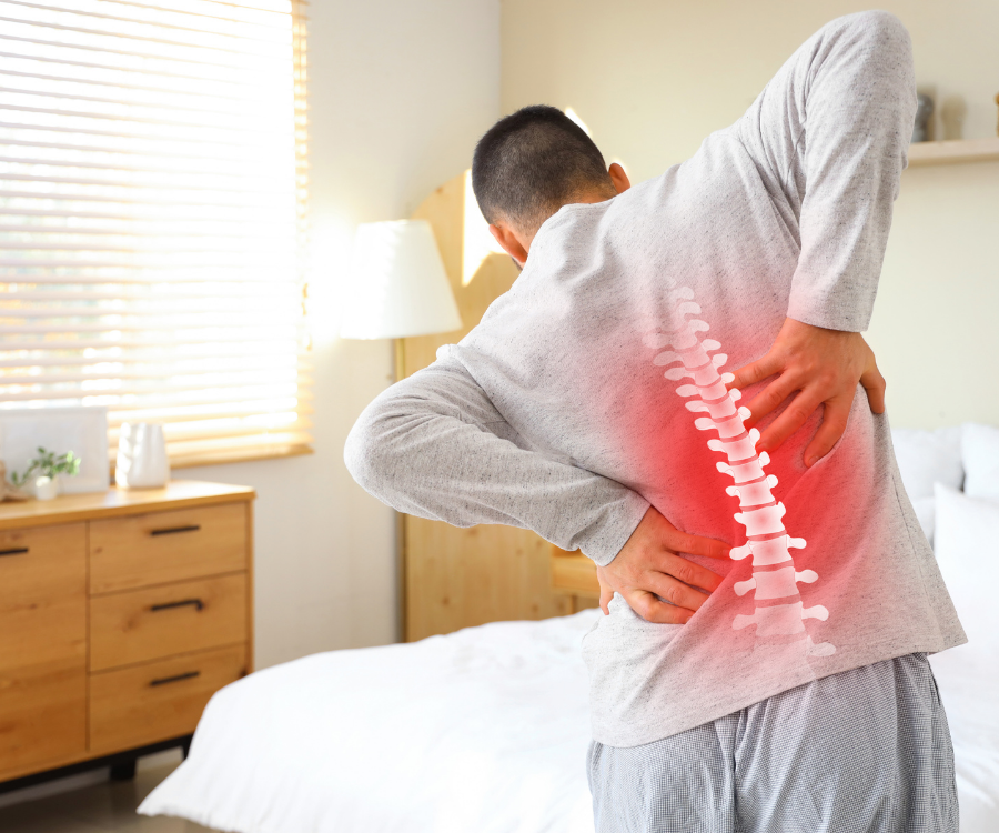
Can an X-Ray Find the Cause of Your Back Pain?
An X-ray can detect bony structural issues—such as fractures, severe arthritis, and spinal alignment problems—but it cannot detect soft-tissue causes like herniated discs, torn ligaments, or nerve compression. For the vast majority of people with acute low back pain, an X-ray will not pinpoint the exact source of discomfort.
How Spinal X-Rays Work
X-rays rely on electromagnetic radiation absorbed at different rates by different tissues. Dense structures like bone absorb radiation readily and appear white or light gray. Soft tissues—muscles, tendons, spinal discs, and nerves—allow radiation to pass straight through, appearing dark or invisible.
Because the spine is a complex mechanical system consisting largely of soft tissues supporting bony vertebrae, standard radiography captures only half the picture.
| What an X-Ray Can Show | What an X-Ray Cannot Show |
| Vertebral compression fractures | Herniated, bulging, or slipped discs |
| Spinal curvature issues (scoliosis, kyphosis) | Sciatica and compressed nerve roots |
| Spondylolisthesis (vertebra slipped forward) | Strained back muscles or sprained ligaments |
| Bone spurs and joint space narrowing (osteoarthritis) | Spinal cord inflammation or tumors |
| Bone erosion from advanced infections or tumors | Sacroiliac (SI) joint soft-tissue dysfunction |
Why Doctors Don’t Always Order an X-Ray Immediately
Clinical guidelines from organizations like the American College of Physicians advise against routine imaging for non-specific, acute low back pain during the first four to six weeks.
- Most acute pain is soft-tissue related: Heavy lifting or awkward twists usually cause ligament sprains or muscle strains. An X-ray reveals nothing about muscle recovery.
- False alarms (“incidental findings”): Natural age-related wear, mild disc narrowing, and minor bone spurs appear on scans of adults who experience zero back pain. Pinning current pain on an incidental bony abnormality often leads to unnecessary procedures, anxiety, and prolonged disability.
- Unnecessary radiation: While spinal X-ray radiation doses are modest, avoiding unnecessary exposure remains sound medical practice.
When Is an X-Ray Actually Necessary?
Physicians evaluate back pain against specific clinical indicators called red flags. An immediate X-ray is warranted when clinical history points to a structural bone compromise:
- Recent major trauma: A car collision, severe fall, or sports impact.
- Minor trauma in high-risk patients: A minor slip or sudden twist in someone with known osteoporosis or extended corticosteroid use.
- Unexplained weight loss or a cancer history: Screening for potential metastatic bone lesions.
- Visible structural deformity: A sudden, noticeable shift in spine curvature or height.
- Persistent pain that fails to improve: Symptoms lasting longer than 6 weeks despite conservative physical therapy and activity modification.
Emergency Note: If severe back pain is accompanied by sudden bowel or bladder incontinence, progressive numbness between the thighs (“saddle anesthesia”), or rapidly worsening leg weakness, seek immediate emergency care. These are signs of cauda equina syndrome, which requires urgent surgical decompression, typically evaluated via emergent MRI rather than X-ray.
X-Ray vs. MRI vs. CT: Choosing the Right Tool
When imaging is indicated, doctors match the diagnostic tool to the suspected underlying pathology.
- X-Ray: Best for rapid, low-cost assessments of bone integrity, gross alignment, and structural stability.
- MRI (Magnetic Resonance Imaging): The gold standard for soft tissues. Uses magnetic fields and radio waves to produce cross-sectional images of the spinal cord, spinal nerves, and intervertebral discs.
- CT (Computed Tomography): An advanced series of computer-combined X-rays that renders highly detailed 3D bony cross-sections, frequently used to assess complex trauma or prepare for surgical hardware placement.
What to Do Instead of Rushing to Imaging
For standard back pain without red-flag symptoms, active recovery is the standard first-line treatment:
- Keep moving gently: Light walking maintains blood circulation and prevents joint stiffness. Prolonged bed rest slows recovery.
- Use heat or cold therapy: Apply ice packs during the first 48 hours to blunt sharp inflammation, then switch to heat to relax spasms.
- Targeted physical therapy: A physical therapist assesses movement mechanics, core activation, and hip mobility to relieve strain on spinal joints without needing an image to start rehabilitation.
No comment