
Hidden Fractures: When an X-Ray Doesn’t Tell the Full Story
You take an awkward tumble on an icy pavement, catch yourself on an outstretched hand, or feel a sudden, sharp ache in your foot halfway through marathon training. At the urgent care clinic, the doctor orders standard radiographs. Twenty minutes later, you receive the verdict: “Good news, your X-rays are completely normal. It’s just a sprain.”
Yet days pass, the throbbing intensifies, weight-bearing feels impossible, and basic mobility remains compromised.
A clean X-ray is often treated as definitive proof of intact bone. In reality, conventional radiography has clear diagnostic limits. In clinical practice, these missed breaks are known as occult fractures—injuries where structural damage exists within the bone architecture, yet remains invisible on initial two-dimensional X-ray projections.
Why X-Rays Miss Fractures
Conventional radiography relies on tissue density. Dense structures like cortical bone absorb X-ray beams effectively, casting white silhouettes onto a detector, while softer tissues allow beams to pass through, appearing dark grey or black.
For a fracture to appear on an X-ray, the bone fragments must be physically separated, displaced, or display a visible gap (a radiolucent line). Several physiological and mechanical factors can mask these injuries:
- Two-Dimensional Limitations: An X-ray flattens complex, three-dimensional skeletal structures into a single plane. If a fracture line runs obliquely or is obscured behind a denser cortical ridge, it will not register on a standard orthogonal view.
- Non-Displaced Microarchitecture: In nondisplaced fractures, hairline fissures, and early stress reactions, the bone fragments remain flush against one another. Without spatial displacement or cortical disruption, the X-ray beam passes through as though the bone were solid.
- The Biological Healing Delay: Bone remodeling takes time. Immediately following an injury, the fracture margins are razor-thin. Over the subsequent 7 to 14 days, osteoclasts resorb damaged micro-bone tissue along the fracture margins, subtly widening the gap. Consequently, a fracture that was invisible on day one frequently becomes obvious on day ten.
High-Risk Areas for Occult Fractures
Certain anatomical regions possess complex vascular arrangements, intricate articular geometry, or heavy biomechanical loading patterns that make them especially susceptible to hidden fractures.
1. The Scaphoid (Wrist)
The scaphoid bone sits below the thumb in the anatomical “snuffbox.” It is the most commonly fractured carpal bone, typically injured when falling onto an outstretched hand (FOOSH). Because of its curved shape and vascular supply—which enters from the distal end and runs backward (retrograde flow)—an undetected scaphoid fracture risks avascular necrosis (bone tissue death) and permanent nonunion.
2. The Femoral Neck (Hip)
In elderly patients with osteopenia or osteoporosis, low-energy falls can crack the femoral neck without displacing the bone. Initial pelvic radiographs may appear pristine. If left unrecognized, continued weight-bearing can cause sudden displacement, converting an easily treatable fracture into an unstable injury requiring total joint replacement.
3. Metatarsals and Tibia (Stress Fractures)
Common in runners, military recruits, and dancers, stress fractures do not stem from a single violent event. They represent fatigue failure caused by repetitive mechanical stress. On initial presentation, standard X-rays miss up to 50% to 70% of early stress fractures, as the initial damage is primarily microscopic trabecular micro-fracturing and bone marrow edema rather than a cortical break.
4. The Pediatric Growth Plate (Salter-Harris Type I)
Children’s bones are capped by cartilaginous growth plates (physis). Cartilage does not contain calcium and appears radiolucent (dark) on X-rays. A shearing injury that cleanly separates through the growth plate without displacing the adjacent bone (Salter-Harris I) leaves zero footprint on an X-ray film.
Comparing Diagnostic Modalities
When physical symptoms point to an injury that an X-ray fails to confirm, secondary imaging modalities provide the necessary resolution:
| Modality | Diagnostic Role | Key Strengths | Limitations |
| Plain Radiography (X-Ray) | First-line triage | Inexpensive, rapid, low radiation dose, detects obvious displacement | Low sensitivity for early stress fractures, marrow lesions, and non-displaced fissures |
| Magnetic Resonance Imaging (MRI) | Gold standard for occult injury | Highly sensitive; detects bone marrow edema, trabecular micro-damage, and soft-tissue injury | Higher cost, longer scan times, availability constraints, contraindications (implants) |
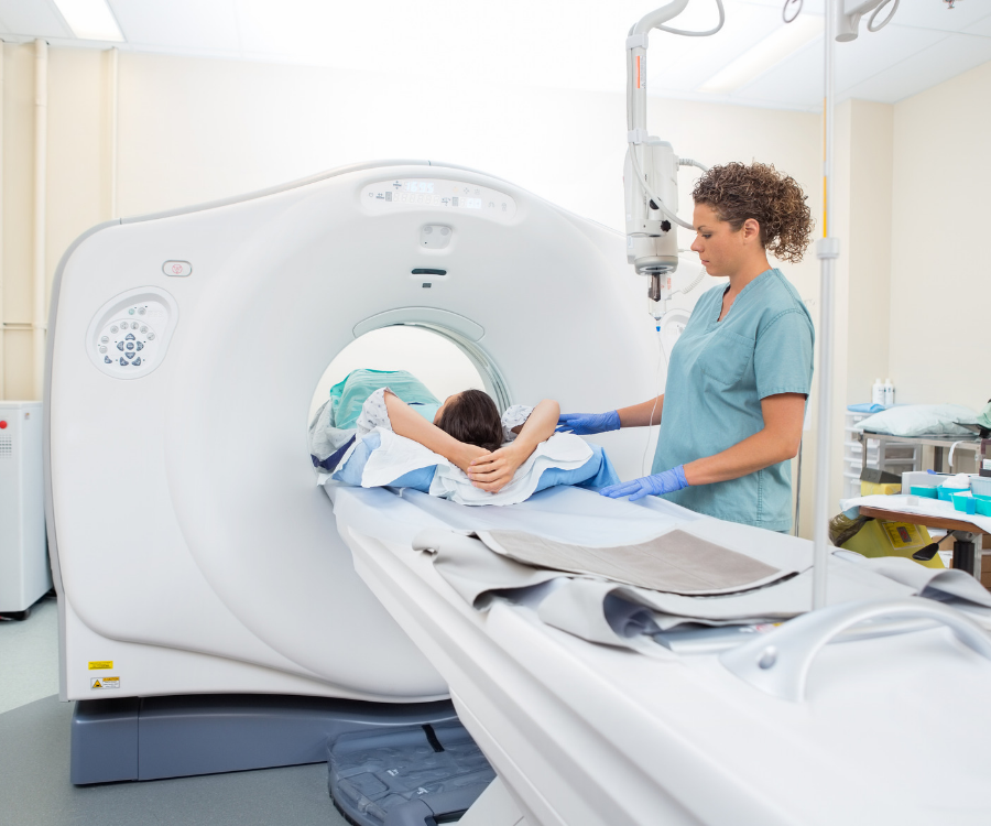
| Computed Tomography (CT) | Multi-planar cortical evaluation | Excellent cross-sectional bone detail; maps complex joint and articular fractures | Ionizing radiation exposure, less sensitive than MRI for pure marrow contusion |
| Musculoskeletal Ultrasound | Superficial cortical assessment | Bedside availability, dynamic evaluation, no radiation | Highly operator-dependent; cannot visualize deep intra-articular structures |
Clinical Red Flags: When to Push for Advanced Imaging
A negative radiograph should never override unmistakable physical findings. If you or a patient encounter the following signs after a traumatic impact or sudden repetitive loading, assume a fracture is present until ruled out:
- Focal, Pinpoint Bone Tenderness: Severe pain elicited by pressing directly on a single bony prominence (e.g., the anatomical snuffbox, the base of the fifth metatarsal, or the pubic ramus).
- Inability to Bear Weight: Inability to take four independent steps immediately following the injury and during the clinical exam.
- Axial Loading Pain: Pain provoked along the shaft of a long bone when gently tapping the heel or compressing the bone lengthwise.
- Disproportionate Swelling and Ecchymosis: Deep, progressive bruising and localized swelling that persists beyond standard soft-tissue recovery timelines.
When these clinical markers are present, standard orthopedic protocol dictates protective immobilization (such as a splint or non-weight-bearing boot) paired with either repeat plain radiography in 10 to 14 days or prompt referral for an MRI or low-dose CT scan.
No comment