
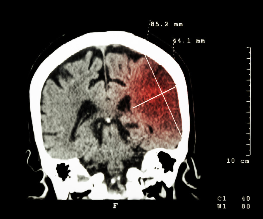
In emergency medicine, the phrase “time is brain” is a clinical reality: during an acute stroke, approximately 1.9 million neurons die every minute blood flow remains disrupted. Computed Tomography (CT) scans serve as the front-line neuroimaging workhorse in emergency departments worldwide, delivering rapid, precise views of intracranial structures to drive life-saving treatment decisions within minutes.
How a CT Scan Identifies a Stroke
A Computed Tomography scan combines narrow X-ray beams with advanced computer algorithms to create cross-sectional “slices” of the skull and brain. Dense structures like bone appear bright white, fluid-filled cavities look black, and brain tissue occupies subtle intermediate shades of gray.
When acute stroke symptoms occur, an emergency CT scan immediately answers the most critical diagnostic question: Is there bleeding in the brain?
- Differentiating Ischemic vs. Hemorrhagic Strokes: Strokes fall into two main categories:
- Ischemic stroke (~87% of cases): A blood clot blocks a cerebral artery, starving brain tissue of oxygen.
- Hemorrhagic stroke (~13% of cases): A weakened blood vessel ruptures, spilling blood directly into brain parenchyma or the subarachnoid space.A standard non-contrast head CT detects acute intracranial hemorrhage almost immediately because extravasated, pooled blood appears bright white.
- Guiding Urgent Thrombolytic Therapy: Clot-busting medications (such as IV Alteplase or Tenecteplase) must be administered within a strict 3-to-4.5-hour window from symptom onset. These drugs are life-saving for ischemic strokes but fatal if administered to someone with a hemorrhage. A clear initial CT ruling out blood is the green light for thrombolysis.
- Spotting Early Ischemic Changes: While early ischemic damage can take several hours to fully demarcate on a plain CT, experienced clinicians look for subtle hallmarks: loss of gray-white matter differentiation, cortical sulcal effacement, and the “hyperdense vessel sign,” where a fresh thromboembolism inside an artery shows up as a bright streak.
- Excluding Stroke Mimics: Brain tumors, subdural hematomas, cerebral abscesses, and trauma can mimic classic stroke signs like sudden facial droop or hemiparesis. A CT scan rules out structural mimics in seconds.
Advanced CT Modalities in Modern Stroke Care
Emergency stroke care has expanded beyond basic scans into multi-modal protocols that map blood flow and vascular anatomy in real time:
| CT Technique | Diagnostic Role | Clinical Value |
| Non-Contrast CT (NCCT) | Baseline screening for acute bleeding, edema, and mass effect. | Rules out hemorrhage within seconds to clear patients for clot-busting therapy. |
| CT Angiography (CTA) | Uses an intravenous iodine contrast agent to visualize arteries from aortic arch to circle of Willis. | Identifies large vessel occlusions (LVOs), aneurysms, or carotid stenosis, qualifying patients for mechanical thrombectomy. |
| CT Perfusion (CTP) | Tracks blood transit times across cerebral tissue beds via dynamic contrast tracking. | Maps the irreversibly damaged “infarct core” against the salvageable “ischemic penumbra”. |
By distinguishing dead brain core from reversibly injured tissue, CT Perfusion enables endovascular thrombectomy—physically retrieving the clot with a stent-retriever—well beyond standard time windows, extending treatment options up to 24 hours after symptom onset in qualified candidates.
CT vs. MRI in Emergency Stroke Diagnosis
| Attribute | Non-Contrast CT Scan | Magnetic Resonance Imaging (MRI / DWI) |
| Acquisition Speed | Under 5 minutes | 15 to 45 minutes |
| Emergency Availability | Ubiquitous in 24/7 emergency rooms | Limited off-hours access in community settings |
| Hemorrhage Detection | Exceptional (hyperdense acute blood) | High (requires dedicated gradient-echo/SWI sequences) |
| Hyperacute Ischemia | Less sensitive in the first 0–3 hours | Pinpoints microscopic ischemia within minutes via DWI |
| Safety Constraints | Low ionizing radiation dose, iodine allergy | No radiation; barred by non-MRI-safe implants/metal |
While MRI Diffusion-Weighted Imaging (DWI) is the most sensitive imaging method for detecting tiny, early ischemic events, CT remains the primary global triage standard because of speed, availability, and instant triage capability.
The Patient Experience: What to Expect During the Scan
Undergoing a head CT is non-invasive and painless:
- Positioning: You lie flat on a motorized gantry table with your head placed comfortably in an ergonomic headrest.
- The Scanner: The table glides into the large, doughnut-shaped gantry opening. Unlike standard MRIs, the scanner ring is open and does not create an enclosed tunnel.
- Contrast Injection (if CTA/CTP ordered): A small IV catheter in the arm or hand delivers an iodinated contrast agent. You may feel a warm flush across your body and a temporary metallic taste, both of which pass within 20 to 30 seconds.
- Duration: The actual image acquisition takes less than two minutes. The entire procedure—from table positioning to image reconstruction—is finished in 5 to 10 minutes.
When to Seek Immediate Medical Evaluation
A CT scan can only save brain tissue if you reach the hospital in time. Remember the B.E. F.A.S.T. acronym to recognize early signs of stroke:
- B – Balance: Sudden loss of balance or coordination.
- E – Eyes: Sudden double vision or partial vision loss.
- F – Face: Facial drooping or an uneven smile.
- A – Arms: Sudden weakness or numbness in one arm or leg.
- S – Speech: Slurred speech, difficulty speaking, or confusion.
- T – Time: Call emergency services (911 or local emergency numbers) immediately.
Never drive yourself or wait to see if symptoms pass. Getting to a CT-equipped acute stroke center as fast as possible remains the single greatest determinant of a complete recovery.
No comment