
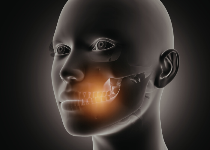
Seeing the Hidden Stress: How X-Rays Reveal Jaw and TMJ Disorders
When you experience a persistent ache in your jaw, a clicking sound when you eat, or the frustrating sensation of your jaw “locking,” the cause isn’t always visible to the naked eye. While a physical exam is the first step, diagnosing Temporomandibular Joint (TMJ) disorders often requires looking beneath the surface.
In the world of modern dentistry and maxillofacial health, X-rays are the primary “detective tools” used to identify what is happening inside your jaw joint. Here is a breakdown of how imaging helps solve the mystery of jaw pain.
What is the TMJ?
The Temporomandibular Joint acts like a sliding hinge, connecting your jawbone to your skull. You have one joint on each side of your face. Because this joint handles complex movements—rotating, gliding, and shifting—it is susceptible to various types of strain, wear, and tear.
Why Your Dentist Needs an X-Ray
A standard visual check can show inflammation or limited movement, but an X-Ray is essential for:
- Checking Bone Alignment: Seeing how the “ball” (condyle) sits in the “socket” (fossa).
- Identifying Arthritis: Spotting bone spurs or the wearing down of the joint surface.
- Ruling Out Other Issues: Ensuring the pain isn’t caused by impacted teeth, infections, or bone cysts.
Common Types of X-Rays for Jaw Problems
Not all X-rays are created equal. Depending on your symptoms, a specialist might use different imaging techniques:
1. Panoramic X-Rays (Orthopantomogram)
This is the most common “overview” film. It provides a broad, ear-to-ear view of your teeth, jawbone, and TMJs. It’s excellent for spotting structural abnormalities or major bone changes, though it provides less detail on the soft tissue (like the cartilage disc).
2. CBCT (Cone Beam Computed Tomography)
For a more advanced look, a CBCT scan provides 3D images. This is the gold standard for looking at bone detail. It allows doctors to see the exact shape of the joint from every angle, making it much easier to spot tiny fractures or subtle erosions caused by TMJ disorders.
3. Tomography
This specific type of X-ray focuses on “slices” of the joint. It is particularly helpful for seeing the joint in different positions—such as when your mouth is fully open versus when it is closed—to see how the joint moves in real-time.
What Doctors Look For on the Film
When a specialist reviews your jaw X-ray, they are looking for specific red flags:
- Flattening of the Joint: A healthy joint is rounded. Flattening often indicates chronic pressure or osteoarthritis.
- Erosion: “Rough” edges on the bone can signal advanced inflammation.
- Joint Space Narrowing: If the space between the bones is too small, it suggests the protective disc has shifted or worn thin.
Moving Beyond the Bone: When X-Rays Aren’t Enough
It’s important to note that X-rays primarily show bone. If your specialist suspects the problem lies with the soft tissue disc (the cushion between the bones) or the muscles, they may recommend an MRI (Magnetic Resonance Imaging). Unlike X-rays, an MRI can “see” the cartilage and help determine if the disc is slipped or damaged.
Summary
If you are struggling with jaw clicking, popping, or pain, an X-ray is often the first step toward a pain-free smile. By capturing a clear picture of your internal anatomy, your dental professional can move away from guesswork and create a targeted treatment plan—whether that involves a custom night guard, physical therapy, or other interventions.
Don’t ignore the ache. A simple scan could be the key to unlocking a more comfortable life.
No comment