When it comes to diagnosing medical conditions, doctors have a range of powerful imaging tools at their disposal. Three of the most common are X-rays, MRI scans, and CT scans. While they all provide valuable insights into the human body, they work in different ways and are best suited for specific purposes. Understanding the differences between these technologies can empower you to have more informed conversations with your healthcare provider.
X-rays: The Quick and Easy Overview
X-rays are the oldest and most widely used form of medical imaging. They use electromagnetic radiation to create images of your bones and dense tissues. Think of it like shining a light through your hand – the bones block more light, creating a shadow image.
- How it works: X-rays pass through the body and are absorbed differently by various tissues. Denser tissues, like bone, absorb more radiation, appearing white on the image. Less dense tissues, like soft tissues, absorb less radiation, appearing darker.
- Best for: X-rays are excellent for visualizing broken bones, detecting foreign objects, and identifying some lung problems. They are also relatively quick and inexpensive.
- Limitations: X-rays provide limited detail of soft tissues and can’t differentiate between different types of soft tissue as effectively as other imaging methods. They also involve a small amount of radiation exposure.
MRI Scans: Detailed Soft Tissue Imaging
Magnetic Resonance Imaging (MRI) uses a powerful magnetic field and radio waves to create detailed images of organs, soft tissues, and bones. It’s like taking a very close look at the body’s internal structures.
- How it works: MRI uses a strong magnetic field to align the protons in your body’s tissues. Radio waves are then emitted, disrupting this alignment. The way the protons realign provides information that is used to create detailed images.
- Best for: MRI scans excel at visualizing soft tissues, making them invaluable for diagnosing conditions affecting the brain, spinal cord, muscles, ligaments, and tendons. They are also used to detect tumors and other abnormalities.
- Limitations: MRI scans are more expensive and time-consuming than X-rays or CT scans. They can also be claustrophobic for some patients, and they are not suitable for people with certain metal implants.
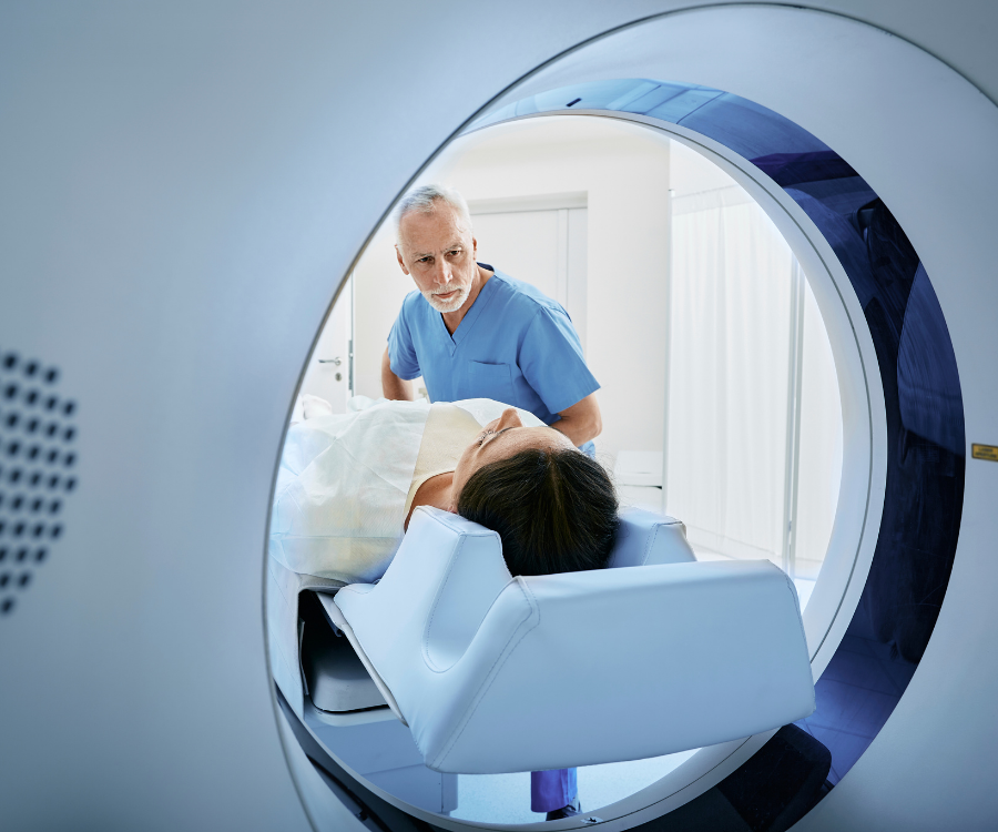
CT Scans: Cross-Sectional Views of the Body
Computed Tomography (CT) scans use X-rays to create cross-sectional images of the body, like slices of bread. This allows doctors to see detailed views of bones, soft tissues, and blood vessels.
- How it works: A CT scanner rotates around the patient, taking multiple X-ray images from different angles. A computer then combines these images to create detailed cross-sectional views.
- Best for: CT scans are particularly useful for visualizing complex bone fractures, detecting internal bleeding, and diagnosing lung conditions. They are also often used in emergency situations.
- Limitations: CT scans involve higher radiation exposure than X-rays, although the risk is still generally considered low. They are also less effective than MRI for visualizing certain soft tissues.
No comment