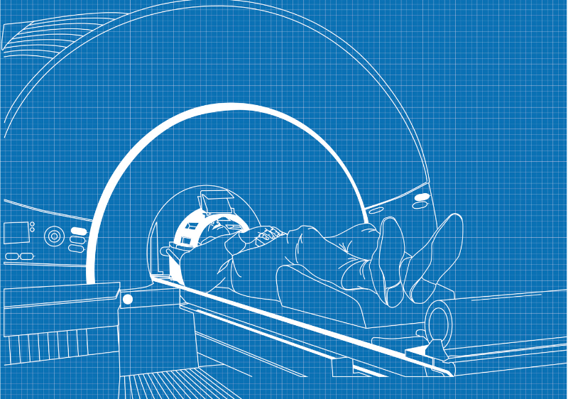
When every second counts, the “gold standard” for stroke imaging is undergoing a massive transformation. For years, the debate was simple: CT for speed, MRI for detail. But in 2026, the lines are blurring.
Thanks to Artificial Intelligence (AI) and “ultrafast” scanning protocols, MRI is no longer the “slow” option. It is becoming a rapid-fire diagnostic powerhouse that can see a stroke in its earliest minutes.
Here is a look at what has improved in MRI for stroke diagnosis and why it matters for patient recovery.
1. Speed: The 3-Minute Stroke Protocol
The biggest historical hurdle for MRI was time. A standard brain MRI used to take 15 to 30 minutes—an eternity when brain cells are dying.
Today, Deep Learning (DL) Reconstruction has changed the game. New “ultrafast” MRI protocols can now capture high-quality diagnostic images in under 3 minutes. These AI-driven scans are “interchangeable” with traditional long-form scans, meaning doctors get the same accuracy in a fraction of the time.
2. AI-Powered Precision
It isn’t just about taking the picture faster; it’s about reading it better. AI algorithms (like those from RapidAI or Viz.ai) now run alongside the MRI scanner to:
- Automatically detect Large Vessel Occlusions (LVOs): Identifying major blockages instantly.
- Quantify the “Penumbra”: AI can pinpoint exactly how much brain tissue is “at risk” but still salvageable versus what is already lost.
- Reduce Human Error: AI acts as a “second pair of eyes” for radiologists, highlighting subtle changes that might be missed in a high-pressure ER environment.
3. Beyond Contrast: Arterial Spin Labeling (ASL)
Traditionally, checking blood flow required injecting a contrast agent (gadolinium). This was a problem for patients with kidney issues or those in a extreme hurry. Arterial Spin Labeling (ASL) is a non-contrast MRI technique that uses the water in the patient’s own blood as a natural tracer. It allows doctors to map cerebral blood flow without needles or dyes, making the process safer and more versatile.
4. Portability: Bringing the MRI to the Patient
We are seeing the rise of Low-Field Portable MRI systems. While these don’t replace the high-resolution 3T magnets found in radiology departments, they can be wheeled directly into intensive care units or even specialized “Stroke Ambulances.”
- Benefit: They allow for continuous monitoring of a patient’s brain without the risk of transporting a critically ill person through a hospital.
5. Better Dating of “Wake-Up” Strokes
About 20% of strokes are “wake-up” strokes, where the patient discovers symptoms upon waking. Because the “time of onset” is unknown, these patients were historically denied life-saving clot-busting drugs. Improved DWI-FLAIR Mismatch imaging allows doctors to “date” a stroke. If the injury shows up on one sequence (DWI) but not yet on another (FLAIR), it’s likely less than 4.5 hours old—opening the door for emergency treatment that would have previously been deemed too risky.
The Bottom Line
MRI is no longer the “second-tier” choice for acute stroke because of its speed. With the integration of AI, non-contrast blood flow mapping, and ultrafast sequences, it provides a “one-stop-shop” for diagnosis, prognosis, and treatment planning.
No comment